
Molecular Breast Imaging (MBI) is an exciting new imaging technology that has great promise for more accurate (and earlier) breast cancer diagnosis. Studies have shown that early detection and treatment make a tremendous difference in outcomes. While mammography is the standard of care in screening, it is less effective in finding tumors in dense breast tissue. So how does MBI help in cancer detection?
- When used in conjunction with mammography screening, the likelihood of tumor detection increases significantly with an MBI exam
- An MBI exam can also rule out cancer more conclusively when a mammogram is indeterminate, which, in turn, can eliminate unnecessary biopsies
FAQ
Following are some commonly asked questions that women have about an MBI exam.
How do I know if I have dense breast tissue?
Contrary to what you might think, breast density does not correlate to how heavy or firm a woman’s breasts are. At present there is no agreed upon method for measuring breast density, but there is agreement that women with dense breast tissue are more likely to develop cancer (studies estimate between 3 and 6 times more likely). Studies have also shown that mammograms are less effective in identifying cancers in dense breasts, because, in mammograms, cancers can appear white like dense breast tissue. While some states in the U.S. have laws requiring that mammography reports include breast density, there are no recommendations or screening guidelines associated with dense breast assessment. You should discuss your breast cancer risk factors with your doctor.
Are there any other reasons for having an MBI?
Your doctor may recommend an MBI if your mammogram was indeterminate (the radiologist has a question about whether there is a breast cancer tumor that the mammogram is not showing clearly). An MBI exam may also be recommended if you cannot tolerate an MRI. The compression is a fraction of what a mammogram requires, so it’s more comfortable than a mammogram.
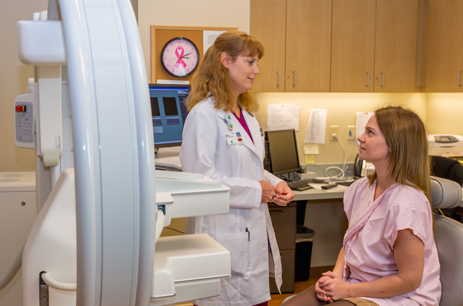
What is the exam like?
There is no advance preparation required for the exam. You will receive a low dose injection of a radiotracer intravenously. The tracer accumulates in cancer cells, and is not influenced by breast density. You can sit comfortably in front of the LumaGEM molecular breast imaging system. Your breast is compressed between two special cameras, which take a series of images. Generally, these will be the same views that were taken during your mammogram to make comparison easier. The procedure takes about 40-45 minutes, and the images are available immediately for the radiologist to review.
Is all this safe? Are there any allergic reactions to the radiotracer? Is it a harmful amount of radiation?
There are no reported side effects from the radiotracer. The amount of radiation is extremely low. You can resume normal activities as soon as the exam is over. The tracer is cleared through natural bodily functions within 6 hours.
Is the exam more likely to show that I have cancer? If the exam does show a lesion, what is the next step?
MBI, in combination with a standard screening mammography, provides much higher sensitivity (91% compared to 27% sensitivity with mammography alone). The likelihood is much higher that an MBI will detect a breast cancer tumor (possibly at an earlier stage) in dense breast tissue if your mammogram is inconclusive or even negative. If your MBI exam reveals a suspicious lesion, then your doctor will likely suggest a biopsy. If your MBI exam confirms that you are cancer-free, it may eliminate an unnecessary biopsy.
I have cancer. Now what?
If you’ve been diagnosed with breast cancer or have a suspicious mammogram, your doctor should refer you for a Breast PET Scan. With the Solo II Breast PET Scanner, doctors can see cancers as small as the width of a grain of rice. These images pinpoint the location of suspicious masses, giving your physician a “map” so they can better select your treatment options and/or surgical plan.
Doctors can better plan to treat breast cancers. This accuracy could result in breast-conserving surgery or lumpectomies. Knowing the exact location and extent of the cancer guides doctors during surgery and helps ensure that they remove all suspicious tissue. Your doctor may also use Breast PET to monitor treatment or to check for a recurrence of disease.
Click here for MBI Addresses Challenges of Dense Breast Tissue by Auntminnie.com